Both times I ended up in hospital, somebody wheeled me off to a scanner and nobody explained why that machine and not a different one. I was too sore to ask. The second time, in Zlín, a CT severity score of 8 out of 10 went into my discharge report, and I only worked out what that number meant months later, at home, reading guidelines I should have been handed on day one.
So here’s what I wish someone had said while I was lying on that trolley. Each scan answers one question. Once you know which question, the order stops feeling random and you can ask better ones yourself.
Ultrasound: are there gallstones?
The ordinary kind. Gel on your belly, a probe pushed up under the ribs, ten minutes, no needles. It’s usually the first scan you get, often within a day of admission.
The question it answers is whether gallstones caused this. The 2024 ACG guideline suggests transabdominal ultrasound in everyone with acute pancreatitis, and a repeat if the first look is inconclusive. Sensitivity for picking up the biliary cause runs around 70 percent, according to the American College of Radiology.
Here’s the part that confused me. Sound doesn’t travel through gas, and an inflamed gut is full of gas, so the pancreas itself is often hidden, especially in severe pancreatitis where the bowel has gone quiet. If your report says the pancreas couldn’t be assessed, the scan still did its job. It was looking at your gallbladder and your bile duct.
CT: how much damage, and is tissue dying?
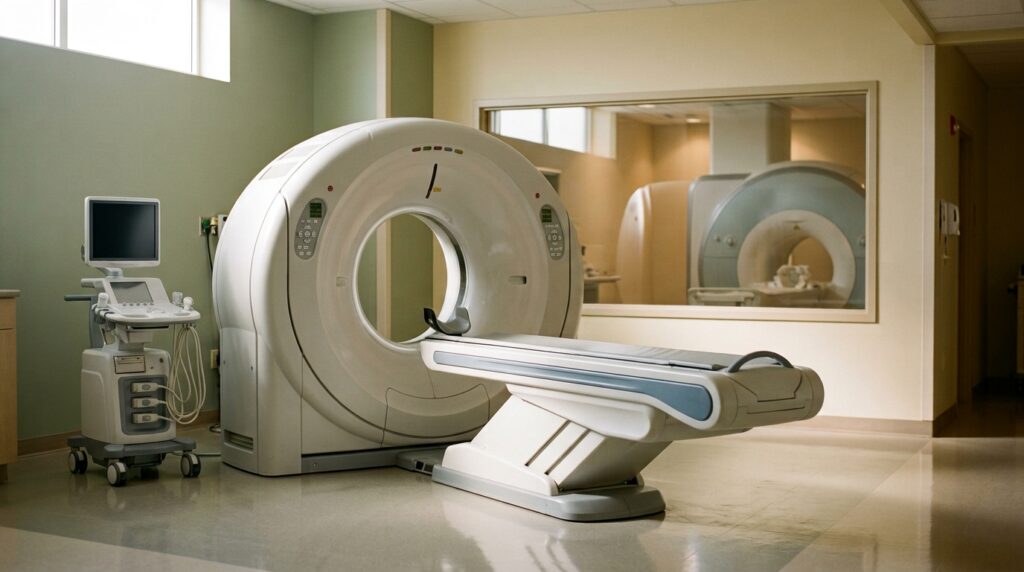
Contrast into a vein, a few minutes in the doughnut, done. This is the scan that produces severity scores like mine.
The timing is the bit nobody explains, and it matters more than the machine. ACG 2024 suggests not doing a routine CT at admission to judge severity, and reserving it for people whose diagnosis is unclear or who aren’t improving after 48 to 72 hours. The reason is physical: dead pancreatic tissue takes days to become visible. The Radiology Assistant puts it bluntly, that severity including necrosis can only be reliably assessed by imaging 72 hours after symptoms start, and the ACR notes that waiting 5 to 7 days gives the best picture of necrosis.
Which means an early CT can look calmer than you actually are. There are published cases of a pancreas enhancing normally on day 1 and showing extensive necrosis on day 3. Same patient, same machine, two days apart.
The costs are real. A CT of the abdomen and pelvis is somewhere around 8 mSv, roughly two to three years’ worth of natural background radiation, and a study taken both before and after contrast, which pancreas protocols often are, can be about double that. Then there’s the iodine contrast itself, which is why they ask about your kidneys before they inject.
MRI and MRCP: what are the ducts doing?
MRCP isn’t a separate machine. It’s an MRI protocol that maps the bile ducts and the pancreatic duct, and it’s usually done in the same session as the rest of the MRI.
No radiation, and it can work without IV contrast, so it’s the one they reach for in pregnancy, in kidney impairment, and with contrast allergy. It also beats CT at telling watery fluid from solid dead debris inside a collection, and that difference decides whether something gets drained or left alone. For bile duct stones it’s more sensitive than ultrasound.
The trade-off is time and stillness. It’s long, loud, and full of breath-holds, and motion wrecks the images, so a very sick or agitated patient gets a CT instead.
There’s a version worth knowing about: secretin-enhanced MRCP. They inject secretin, a hormone that makes your pancreas pour out fluid, the ducts fill and stretch, and small branches you couldn’t see become visible. It’s used for pancreas divisum, duct strictures and chronic pancreatitis questions. It adds at least 15 minutes, and side effects like nausea or flushing show up in about 0.5 percent of people.
Endoscopic ultrasound: the one that finds the small stuff
This is the odd one, and the one most patients have never heard of. You’re sedated, a scope goes down through your mouth into the stomach and duodenum, and there’s an ultrasound probe on its tip. The probe ends up a centimetre from your pancreas with only the stomach wall in between. No gas in the way, no fat to shoot through.
The question it answers is what everything else missed. A 2025 systematic review and meta-analysis in Digestive Diseases and Sciences pooled 8 studies comparing EUS with MRCP in people whose pancreatitis had no known cause. EUS found a likely cause about twice as often overall, and it was close to four times better at catching biliary causes: small stones, microlithiasis, sludge. MRCP looked better for pancreas divisum, a duct variant you’re born with, though that particular difference didn’t reach statistical significance, so treat the two as complementary. The pooled studies also varied a lot between themselves, and this is one recent analysis rather than a guideline position.
EUS can also take a tissue sample with a fine needle during the same procedure, which no scanner can do.
If you’ve been told your pancreatitis is idiopathic, meaning cause unknown, this is the test to ask about by name.
ERCP: a treatment that gets mistaken for a scan
ERCP looks like EUS from the outside. Scope, sedation, same room. It’s a different thing. There’s a camera, dye injected into the bile duct, X-rays, and tools that can cut the sphincter and pull a stone out. It treats.
ACG 2024 is specific. If pancreatitis comes with cholangitis, an infected bile duct, ERCP within 24 hours lowers the odds of dying. Without cholangitis or jaundice, when a duct stone is only suspected, you screen with MRCP or EUS first, and diagnostic ERCP should be avoided.
The reason is uncomfortable: ERCP itself causes pancreatitis. European and American endoscopy societies put it at roughly 3 to 10 percent of procedures, about 8 percent in average-risk patients and around 15 percent in high-risk ones. That’s the whole argument for looking with something harmless first. If ERCP is proposed and nobody mentions what they plan to remove or stent, that’s the moment to ask.
What about everything else
A plain abdominal X-ray tells you little about the pancreas. It gives nonspecific findings, and it’s usually ordered to answer a different question, like ruling out a hole in the gut. It can show calcification in a long-damaged pancreas, so it isn’t ordered for nothing.
Blood tests aren’t imaging, but they’re the actual diagnosis. Acute pancreatitis is diagnosed on 2 of 3 things: the typical pain, lipase or amylase at three times the upper limit, or imaging that shows it. So the pain and the lipase alone are enough, which is why a doctor can be certain about your diagnosis before you’ve been near a scanner. You’ll still get the ultrasound, because that one is hunting for the cause, not confirming the diagnosis.
Later on there’s often a repeat CT or MRI, and that’s usually about collections: fluid or dead tissue that’s walled itself off, and whether it needs draining. Interval scans in the weeks after an attack are normal, not a sign that something has gone wrong.
One more worth knowing, because people expect a scan to answer it: enzyme insufficiency, the reason you might end up on Creon, is diagnosed from a stool sample (faecal elastase), not from any picture.
And for chronic pancreatitis the sequence shifts. ACG guidelines put CT or MRI first, where calcification and a dilated duct show up once the damage is established. EUS and secretin MRCP come in when those are inconclusive and the suspicion is still there, because they see earlier changes. EUS is sensitive but not very specific in early disease, meaning it can call chronic pancreatitis in people who don’t have it, which is why it isn’t the opening move.
Four questions worth asking
- Which question is this scan meant to answer?
- Will the result change what you do for me this week?
- Is there contrast, and have my kidneys been checked?
- If no cause has been found, when do I get an EUS?
I asked none of these the first time. I asked most of them the second time, and the second admission was easier to be a patient in, partly for that reason.
Stepan’s Rule: an early CT shows you day one. Necrosis shows up later. Judge that first week by how you feel and what your bloods do, and if the picture and the patient disagree, ask when they plan to scan again.
I’m a patient, not a doctor. Which scan you need and when is a decision for the team holding your chart. What I can tell you is that asking what a scan is for changed how those conversations went for me.
If you’re waiting on a scan right now and reading reports you don’t understand, the PancreasZen community is full of people who’ve read the same sentences. Come in and ask.
Where this comes from
ACG Clinical Guideline: Management of Acute Pancreatitis (2024), for the ultrasound, CT timing and ERCP recommendations. ACR Appropriateness Criteria for acute pancreatitis and RadiologyInfo.org, for sensitivity figures, radiation dose and the limits of each modality. ESGE and ASGE guidance on ERCP-related adverse events. The 2020 ACG chronic pancreatitis guideline. The Radiology Assistant’s acute pancreatitis chapter, for the 72-hour rule on assessing necrosis. A systematic review and meta-analysis in Digestive Diseases and Sciences (published online 2025), for the EUS versus MRCP comparison. The AJR expert panel review on secretin-enhanced MRCP. Percentages are pooled study figures, not promises about your scan.
The PancreasZen book has the questions worth taking to each appointment, so you are not trying to remember them under pressure.